Can you have a mammogram with breast implants?
Yes, you can and should have a mammogram with breast implants if you are in a screening age group or have symptoms — just tell the radiographer about your implants before the examination starts.
Breast implants do not stop you having breast screening, and having implants does not change your underlying risk of breast cancer. What changes is the technique. Implants are dense on X-ray and can hide part of the breast tissue, so the radiographer takes extra images and positions the breast differently to see as much tissue as possible.
A mammogram with implants usually takes a little longer than a standard one, and some screening services book a longer appointment or a specific location for women with implants. You may be asked to sign a form confirming you understand that the implant can hide some tissue and that, very rarely, compression could affect the implant.
The examination applies to women who have had cosmetic breast augmentation and to women with an implant in a breast that still contains natural breast tissue. After a mastectomy with reconstruction, the situation is different, as explained further down.
Having implants is not a reason to skip a screening invitation or to delay getting a new breast symptom checked.
What should I tell the radiographer before a mammogram with implants?
Before your mammogram, tell the radiographer that you have implants, roughly when and where they were placed, whether they are in front of or behind the muscle, and about any symptoms or problems such as firmness or a change in shape.
Useful information to bring or mention includes:
- That you have implants, ideally when you book the appointment as well as on the day.
- The year of surgery and, if known, the implant type (silicone or saline) and manufacturer, for example from your implant card.
- Whether the implants sit beneath the breast tissue or beneath the chest muscle.
- Any previous implant surgery, capsular contracture or known rupture.
- Any new lump, swelling, pain or change you have noticed.
- Any previous breast surgery for cancer or benign lumps, and any radiotherapy.
If you have symptoms, it is usually better not to wait for a routine screening appointment. See your GP or family doctor, who can refer you to a breast clinic for a diagnostic assessment rather than a screening test.
What are Eklund (implant displacement) views?
Eklund views, also called implant displacement views, are extra mammogram images in which the radiographer gently pushes the implant back towards the chest wall and pulls the breast tissue forward, so that more of the breast tissue can be compressed and seen clearly.
A mammogram with implants usually includes:
- Standard views with the implant included, taken with lighter compression, to show the implant and the tissue around it.
- Implant displacement (Eklund) views, in which the implant is moved out of the way and only the breast tissue is compressed.
This means you may have four images per breast rather than the usual two. Displacement views work best when the implant lies beneath the muscle and the breast tissue moves freely over it. They are more difficult when there is capsular contracture, because the firm capsule makes the implant hard to move; in that case the radiographer does what is possible and the radiologist may suggest additional imaging.
The technique is named after the radiologist who described it. Most breast imaging units with experience of implants use it routinely.
Does implant position affect how well a mammogram works?
Yes, implants placed beneath the chest muscle usually allow more breast tissue to be seen on a mammogram than implants placed directly beneath the breast tissue.
Silicone and saline implants both block X-rays, so any tissue directly in front of or behind them can be partly hidden. When the implant is beneath the muscle (submuscular or dual-plane), the breast tissue lies on top of the muscle and is easier to pull forward for displacement views. When the implant sits directly beneath the breast tissue (subglandular), more tissue may overlap the implant.
Other factors that affect image quality include:
- The size of the implant compared with the amount of natural breast tissue.
- Capsular contracture, which makes the implant less mobile.
- Scar tissue or calcium in the capsule around older implants.
- Previous fat grafting, which can leave small oil cysts or calcifications that radiologists are usually able to recognise.
Even with careful technique, a small proportion of breast tissue may not be fully visible. This is one reason why breast awareness and prompt reporting of any change remain important for women with implants. You can read more about the condition that causes firm implants on our capsular contracture page.
- Implant
- Pectoralis muscle
- Ribs
- Muscle cover
Can a mammogram rupture a breast implant?
A mammogram very rarely damages or ruptures a breast implant, and the benefit of screening is generally considered to outweigh this small risk.
Radiographers use less compression over the implant itself and rely on displacement views to compress the breast tissue. The risk is thought to be higher with older implants that are already weakened, with severe capsular contracture, or if an implant is already damaged without anyone knowing.
Some practical points:
- Mammography can feel more uncomfortable with implants; tell the radiographer if it hurts.
- If you notice a change in shape, a new lump, pain or swelling after a mammogram, contact your doctor.
- Saline implant rupture is usually obvious, as the breast deflates over days. Silicone rupture is often “silent”, with no symptoms, and is usually found on ultrasound or MRI.
A mammogram is not a reliable test for silicone implant rupture. If rupture is suspected, ultrasound or MRI is usually used instead.
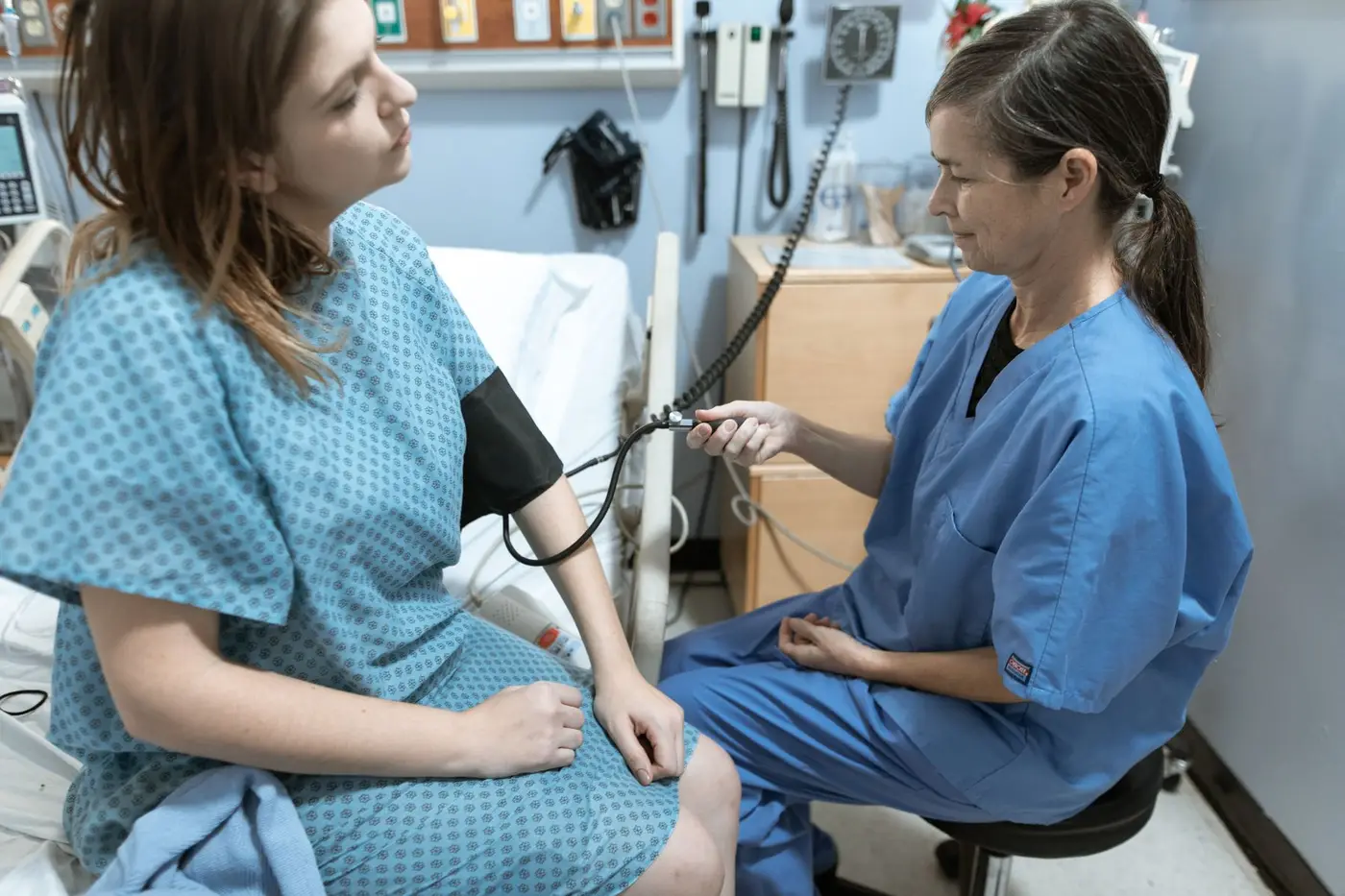
Do I need an MRI or ultrasound to check my breast implants?
Ultrasound and MRI are the main tests for checking whether a silicone implant has ruptured, and some regulators, such as the US Food and Drug Administration (FDA), recommend periodic imaging for silicone implants even without symptoms.
The FDA’s labelling recommendations for silicone gel implants suggest that women without symptoms have an ultrasound or MRI about five to six years after surgery and then every two to three years, and that an MRI is used if there are symptoms or an unclear ultrasound result. Recommendations differ between countries: in the UK and much of Europe, routine imaging of implants without symptoms is generally not part of standard practice, and imaging is arranged when there are symptoms or concerns. Ask your surgeon what is appropriate for you.
Ultrasound
Ultrasound is widely available, quick and uses no radiation. It can often show rupture, fluid around the implant and enlarged lymph nodes, but it depends on the operator and may miss some ruptures.
MRI
MRI without contrast is the most accurate test for silicone implant rupture. MRI with contrast is a different scan, used to look at breast tissue for cancer in certain situations, for example in women at high risk or when other tests are unclear.
These scans are tools for checking the implant. They do not replace mammography for breast cancer screening in women who are of screening age.

Mammogram, ultrasound or MRI: which test is used for what?
A mammogram is used to screen for breast cancer, ultrasound is used to check lumps and often the implant, and MRI is the most accurate test for silicone rupture and is also used for high-risk screening.
| Test | Main purpose with implants | Strengths | Limitations |
|---|---|---|---|
| Mammogram | Breast cancer screening and assessing symptoms | Detects small cancers and calcifications | Implant can hide some tissue; not reliable for silicone rupture |
| Ultrasound | Checking lumps, fluid, lymph nodes and implant integrity | No radiation, quick, widely available | Operator-dependent; may miss some ruptures |
| MRI without contrast | Checking silicone implant rupture | Most accurate test for rupture | Expensive, less available, not tolerated by everyone |
| MRI with contrast | High-risk screening or unclear findings | Very sensitive for breast cancer | More false alarms; only used in selected women |
If a test shows something that needs a closer look, the next step may be further imaging or a needle biopsy, decided by the breast clinic. You can read about these steps on our breast cancer diagnosis page.

Do I need mammograms after breast reconstruction?
After a mastectomy and breast reconstruction, the reconstructed breast is generally not routinely screened with mammograms, but the other breast usually continues to be screened, and you should follow the advice of your breast team.
A mastectomy removes most of the breast tissue, so there is very little tissue left for a mammogram to examine in a reconstructed breast, whether it was rebuilt with an implant or with your own tissue. Follow-up of the reconstructed side usually relies on clinical examination and imaging of any new lump or change.
The situation is different when breast tissue remains:
- After breast-conserving surgery, with or without oncoplastic reshaping, the treated breast usually has regular mammograms as part of follow-up.
- The opposite, untreated breast usually continues to have mammograms, including if it has had an implant or lift for symmetry.
- After fat grafting, oil cysts and small calcifications can appear on imaging; radiologists can usually tell these apart from worrying changes, but tell them about the grafting.
Follow-up schedules differ between countries and between patients, depending on the type of cancer and treatment. Your oncology team decides which imaging you need and how often; see also our page on life after breast cancer.
How does breast screening work with implants in Germany, Bulgaria and the UK?
In Germany, Bulgaria and the UK, women with breast implants can usually take part in breast screening in the same way as other women, but they should mention their implants when booking and at the appointment.
Germany
The national Mammographie-Screening-Programm invites women aged 50 to 75 for a mammogram every two years. Women with implants can take part; tell the screening unit about your implants. For symptoms or questions, the usual first contact is your Frauenarzt or Frauenärztin (gynaecologist), who can arrange diagnostic imaging or referral to a certified breast centre (Brustzentrum).
Bulgaria
In Bulgaria, access to mammography is usually arranged through your GP (общопрактикуващ лекар), who can refer you to a specialist in mammology or breast imaging. Organised screening arrangements and campaigns vary, so ask your GP what is available to you and mention your implants when the examination is booked.
United Kingdom
The NHS Breast Screening Programme invites women aged about 50 to 71 every three years. Women with implants can be screened; the NHS advises telling the screening unit beforehand, as some units arrange appointments for women with implants at a particular site or time.
In all three countries, screening is for women without symptoms. If you notice a change, do not wait for an invitation — see your doctor.

How do I check my breasts if I have implants?
Checking your breasts with implants is similar to checking natural breasts: get to know how they normally look and feel, including the edges of the implant, and report any new change to your doctor.
Implants can make the breast feel different. You may be able to feel the edge of the implant, especially at the sides or lower part of the breast, or small folds called ripples. These are usually normal for you if they have been there since surgery.
When checking, look and feel for:
- A new lump or thickening in the breast, the armpit or around the collarbone.
- Sudden swelling or enlargement of one breast, even months or years after surgery.
- A change in shape, firmness or position of the implant.
- Skin dimpling, redness, a rash or an orange-peel texture.
- Nipple changes or discharge.
- Persistent pain in one area.
Most changes in women with implants are not cancer. Common explanations include benign lumps, folds in the implant or scar tissue; see our page on benign breast lumps. Even so, every new lump should be checked by a doctor.

What happens if a mammogram or self-check finds something?
If a mammogram or self-check finds something unusual, you will usually be offered further imaging and, if needed, a needle biopsy, and most findings in women with implants turn out to be benign.
Possible findings and their usual next steps include:
- A breast lump or area of concern — assessed with ultrasound and further mammogram views, and a biopsy if needed. Biopsies can usually be done safely with implants, using ultrasound guidance to avoid the implant.
- Silicone outside the implant or a suspected rupture — usually confirmed with ultrasound or MRI, and discussed with a plastic surgeon.
- Fluid around the implant (late seroma) — a new swelling appearing a year or more after surgery should be assessed with ultrasound and the fluid tested, because rarely it can be a sign of BIA-ALCL, a lymphoma linked mainly to textured implants.
- Calcifications in the capsule or oil cysts — often benign changes related to older implants or fat grafting.
Waiting for results can be stressful. Breast clinics aim to give results as quickly as possible, and a recall after screening does not mean you have cancer.
How a plastic surgeon can help
A plastic surgeon does not perform or interpret mammograms, but can help when imaging suggests an implant problem such as rupture, capsular contracture or fluid around the implant, and when implants need to be removed or replaced.
Breast screening and the assessment of lumps belong with your GP, gynaecologist, breast clinic and radiologist. Where the findings involve the implant itself, Dr. Fatih Kılıç can:
- Review your ultrasound or MRI reports and explain what they mean for the implant.
- Discuss whether an implant needs to be replaced, removed, or simply monitored.
- Explain options such as implant exchange, capsulectomy, implant removal with a lift, or fat transfer — see breast implant removal and exchange.
- Advise on choices that may make future imaging easier, such as implant position and size, when planning new surgery.
If you have had breast cancer, any implant surgery is planned together with your oncology team, who remain in charge of your cancer follow-up.
Implant concerns after screening: an opinion for patients from Germany, Bulgaria and the UK
Breast screening and the assessment of any lump should always take place with your own doctors at home — your Frauenarzt or breast centre in Germany, your GP and mammology specialist in Bulgaria, or the NHS in the UK. If your mammogram, ultrasound or MRI report mentions an implant problem such as rupture, capsular contracture or fluid around the implant, you can share the reports and photographs online for an opinion from Dr. Fatih Kılıç on implant exchange or removal. Istanbul is about 1 hour 15 minutes by air from Sofia and about 3 hours from major German airports. After any treatment you receive a written summary for your doctor at home. See international patients or contact us.

Frequently asked questions
Can you have a mammogram if you have breast implants?
Yes. Women with breast implants can and should have mammograms when they are of screening age or have symptoms. Tell the radiographer about your implants so that extra implant displacement views can be taken.
Do I need to tell the screening service that I have implants?
Yes. Mention your implants when you book and again on the day. Some services arrange a longer appointment or a particular site for women with implants, and the radiographer will adapt the technique.
Can a mammogram burst a breast implant?
This is very rare. Radiographers use lighter compression over the implant and rely on displacement views. The risk may be slightly higher with old or already damaged implants, so report any change in shape, pain or swelling afterwards.
Is a mammogram more painful with implants?
It can be a little more uncomfortable for some women, especially if there is capsular contracture. Let the radiographer know if it hurts, as they can adjust the compression.
What are Eklund views?
Eklund views, or implant displacement views, are extra mammogram images in which the implant is pushed back against the chest wall and the breast tissue is pulled forward. This lets more breast tissue be compressed and seen clearly.
Can implants hide breast cancer on a mammogram?
Implants can hide part of the breast tissue on a mammogram, which is why extra views are taken. Even then, a small amount of tissue may not be fully visible, so it is important to report any new lump or change to your doctor.
Can a mammogram detect implant rupture?
A mammogram may sometimes show a saline rupture or silicone outside the implant, but it is not a reliable test for silicone rupture. Ultrasound or MRI is usually used to check whether a silicone implant is intact.
How often should silicone breast implants be checked with MRI?
The US FDA suggests ultrasound or MRI about five to six years after surgery and then every two to three years for silicone implants, even without symptoms. Advice differs between countries, and in many European countries imaging is usually arranged when there are symptoms. Ask your surgeon what is right for you.
Do I need mammograms after a mastectomy and reconstruction?
The reconstructed breast is generally not routinely screened with mammograms, because most of the breast tissue has been removed. The other breast usually continues to be screened, and your oncology team will advise on your follow-up.
Does having breast implants increase the risk of breast cancer?
Breast implants do not increase the risk of ordinary breast cancer. Textured implants have been linked to a rare lymphoma called BIA-ALCL, which is a cancer of the immune system rather than of the breast tissue.
At what age does breast screening start in Germany, Bulgaria and the UK?
In Germany, the national programme invites women aged 50 to 75 every two years. In the UK, the NHS invites women aged about 50 to 71 every three years. In Bulgaria, mammography is usually arranged through your GP, so ask your GP what is available to you.

Medically reviewed by Dr. Fatih Kılıç, Plastic, Reconstructive and Aesthetic Surgeon. This page is for general information and does not replace an individual consultation.