How is breast cancer diagnosed?
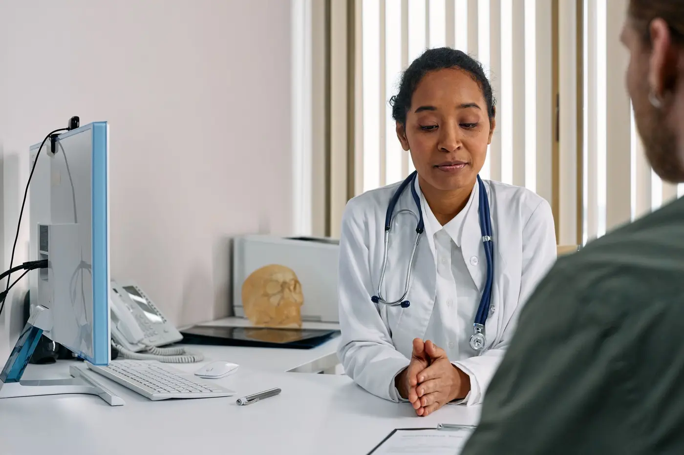
Breast cancer is diagnosed with a combination of a clinical examination, breast imaging and a biopsy, an approach often called the triple assessment. No single test is enough on its own: a scan can raise suspicion, but only a tissue sample examined under a microscope can confirm whether a change is cancer.
Most people come to a breast clinic for one of two reasons: they have noticed a change such as a lump, skin dimpling or nipple discharge, or a routine screening mammogram has shown something that needs a closer look. If you have noticed a change, our guide to breast cancer symptoms explains what to look out for.
Most breast lumps and abnormal mammograms turn out not to be cancer. Even so, every new lump or change should be checked by a doctor promptly, rather than waiting to see if it settles.
The three parts of the triple assessment are:
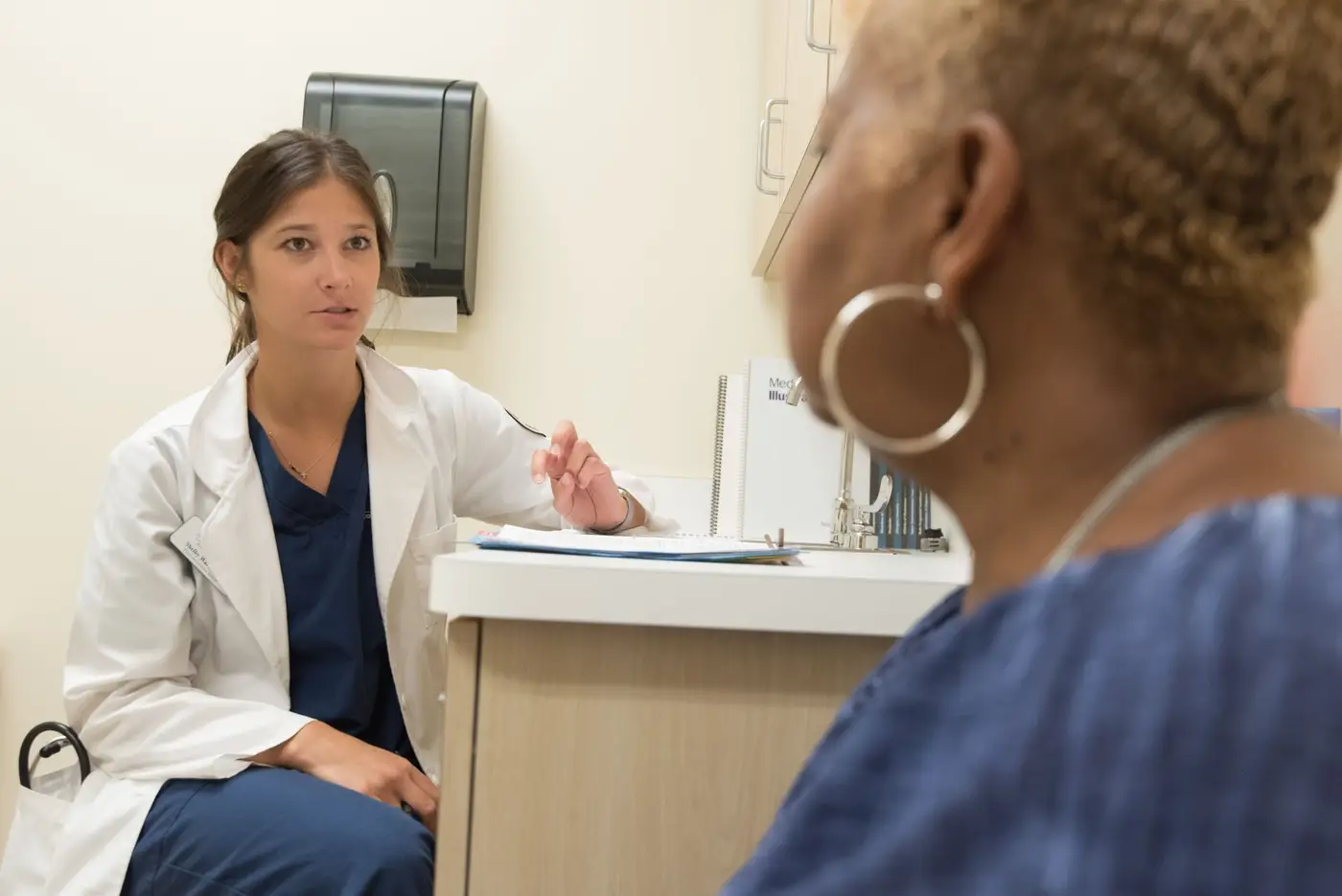
- Clinical examination — a doctor or specialist nurse asks about your symptoms, family history and medicines, and examines both breasts and the lymph nodes in the armpits and above the collarbones.
- Imaging — usually a mammogram and/or a breast ultrasound, sometimes an MRI.
- Biopsy — a small sample of tissue is taken from the area of concern, usually with a needle under local anaesthetic, and sent to a pathologist.
In many specialist breast units these steps can happen at a single visit, often called a one-stop clinic. The final results are then discussed by a multidisciplinary team (MDT) of breast surgeons, radiologists, pathologists, oncologists and breast care nurses before a treatment plan is agreed with you.
What does a mammogram show?
A mammogram is a low-dose X-ray of the breast that can show lumps, areas of distortion and tiny calcium deposits (microcalcifications), some of which may be an early sign of cancer. It is the main test used in breast screening programmes and is usually the first scan for women over about 40 who have symptoms.
During a mammogram, each breast is gently compressed between two plates for a few seconds while the images are taken. This can be uncomfortable, but compression keeps the dose low and the picture sharp. Two views of each breast are usually taken.
What is 3D mammography (tomosynthesis)?
Digital breast tomosynthesis, often called 3D mammography, takes a series of images from different angles and builds them into thin slices. This can make it easier to see through overlapping tissue, particularly in dense breasts, and may reduce the number of women called back for further tests. It is widely used in some countries and in selected situations in others; it is not yet the standard screening test everywhere.
Why are dense breasts harder to read?
Breasts with more glandular and fibrous tissue look white on a mammogram, and so do many cancers. Dense breast tissue is common and normal, especially in younger women, but it can hide small changes. In these cases your team may add an ultrasound or, occasionally, an MRI.
Why might I need a breast ultrasound?
A breast ultrasound uses sound waves to show whether a lump is a fluid-filled cyst or a solid area, and it is often the first test for women under about 40, during pregnancy or breastfeeding, and for many men. It involves no radiation and is painless.
Ultrasound is also used to look at the lymph nodes in the armpit (the axilla) and to guide a biopsy needle precisely into a small lump. If a node looks unusual, a sample may be taken from it at the same appointment. You can read more about why the armpit nodes matter in our guide to breast cancer types and stages.
Simple cysts are very common and are not cancer. If a lump is solid, the radiologist will describe its shape and edges, which helps decide whether a biopsy is needed.
When is a breast MRI used?
A breast MRI uses a strong magnet and a contrast injection to create very detailed pictures, and it is used in selected situations rather than for everyone. It is not usually the first test for a new lump.
Your team may suggest an MRI:
- to measure the size and extent of a cancer more accurately, especially invasive lobular cancer, which can be hard to see on a mammogram;
- when the breasts are very dense or mammogram and ultrasound results do not match;
- to check the other breast in some newly diagnosed patients;
- to monitor the response to chemotherapy given before surgery;
- for screening women at high inherited risk, such as BRCA carriers (see hereditary breast cancer and BRCA);
- to assess breast implants or to look for a hidden primary cancer when cancer has been found in an armpit node.
MRI is very sensitive, which means it sometimes picks up harmless areas that then need further tests or a biopsy. Your team will weigh this against the benefit in your individual situation.
What do BI-RADS categories mean?
BI-RADS (Breast Imaging Reporting and Data System) is a standard scoring scale from 0 to 6 that radiologists use to summarise how likely a mammogram, ultrasound or MRI finding is to be cancer. In the UK a similar 1–5 scale (for example M1–M5 for mammography and U1–U5 for ultrasound) is often used instead.
| Category | What it means | Usual next step |
|---|---|---|
| BI-RADS 0 | Incomplete — more imaging needed | Further views, ultrasound or comparison with old scans |
| BI-RADS 1 | Negative — nothing found | Routine screening |
| BI-RADS 2 | Benign finding, such as a simple cyst | Routine screening |
| BI-RADS 3 | Probably benign (cancer risk generally under 2%) | Short-interval follow-up scan, often at 6 months |
| BI-RADS 4 | Suspicious (subdivided 4A, 4B, 4C from low to moderate-high suspicion) | Biopsy |
| BI-RADS 5 | Highly suggestive of cancer | Biopsy |
| BI-RADS 6 | Cancer already confirmed by biopsy | Treatment planning |
A BI-RADS 4 or 5 result means a biopsy is recommended; it is not a diagnosis. Many BI-RADS 4 findings, particularly 4A, turn out to be benign.
What types of breast biopsy are there?
A breast biopsy removes a small sample of tissue or cells so that a pathologist can look at it under a microscope, and it is the only way to confirm a breast cancer diagnosis. Most biopsies are done with a needle under local anaesthetic, guided by ultrasound, mammography or MRI.
Core needle biopsy
A core needle biopsy is the most common type. A hollow needle takes several thin cores of tissue, each about the size of a grain of rice or a little larger. It gives enough tissue to tell whether a cancer is invasive or in situ and to test its receptors (ER, PR and HER2).
Vacuum-assisted biopsy
A vacuum-assisted biopsy uses a slightly larger needle with gentle suction to collect more tissue through a single small cut. It is often used for microcalcifications seen only on a mammogram (a stereotactic biopsy), for small areas, or to remove some benign lumps completely.
Fine needle aspiration (FNA)
Fine needle aspiration uses a very thin needle to draw out cells or fluid. It is useful for draining cysts and for sampling armpit lymph nodes, but it collects cells rather than a tissue core, so it usually cannot show whether a breast cancer is invasive. For a breast lump, a core biopsy is generally preferred.
After a needle biopsy, a tiny metal marker clip is often left in the breast so the area can be found again later — for example during surgery. Bruising and soreness are common for a few days. Surgical (excision) biopsy is now needed only occasionally.

How do I read a breast cancer pathology report?
A breast pathology report describes what the cancer cells look like and how they behave, and these details guide almost every treatment decision. Your team should go through it with you; it is normal to find the language difficult, so ask them to explain anything unclear.
Invasive or in situ?
In situ means the abnormal cells are still contained inside the milk ducts or lobules — for example ductal carcinoma in situ (DCIS). Invasive means the cells have grown through the duct or lobule wall into surrounding breast tissue, which gives them the potential to spread. The most common invasive types are invasive ductal (no special type, NST) and invasive lobular cancer.
Grade (1, 2 or 3)
The grade describes how different the cancer cells look from normal cells. Grade 1 cells look most like normal cells and tend to grow slowly; grade 3 cells look very different and tend to grow faster. Grade is not the same as stage.
Hormone receptors (ER and PR)
ER (oestrogen receptor) and PR (progesterone receptor) show whether the cancer uses hormones to grow. Most breast cancers are ER-positive, which means hormone (endocrine) therapy is likely to be an important part of treatment.
HER2 status, including HER2-low
HER2 is a protein that helps cells grow. It is tested with a stain scored 0, 1+, 2+ or 3+, sometimes followed by a gene test (ISH or FISH). HER2-positive (3+, or 2+ with a positive gene test) means targeted anti-HER2 drugs are usually recommended. HER2-low (1+, or 2+ with a negative gene test) is a newer category: it matters mainly because some newer drugs may be an option if the cancer ever spreads.
Ki-67
Ki-67 estimates the percentage of cells that are actively dividing. A higher Ki-67 suggests faster growth. Laboratories measure it differently, so it is interpreted alongside the other results rather than on its own.
Size, lymph nodes and margins
After surgery, the report also gives the tumour size, whether any lymph nodes contained cancer and the margins — the rim of healthy tissue around the removed cancer. “Clear” or “negative” margins mean no cancer cells were found at the edge; if cancer reaches the edge, further surgery is sometimes advised. The report may also mention lymphovascular invasion (cancer cells seen in small vessels).

Will I need scans to check if the cancer has spread?
Not everyone needs staging scans: many people with small, early breast cancers and no worrying symptoms do not need body scans, because the chance of finding spread is very low. Scans are usually recommended when the cancer is larger, the lymph nodes are involved, the biology is more aggressive or there are symptoms such as bone pain.
Scans that may be used include:
- CT scan of the chest, abdomen and pelvis to look at the lungs, liver and lymph nodes;
- bone scan to look for changes in the bones;
- PET-CT, which uses a mildly radioactive sugar to highlight active areas, in selected cases — for example locally advanced or inflammatory breast cancer, or when other scans are unclear;
- occasionally an MRI of the brain or spine if there are relevant symptoms.
Blood tests are also done to check general health, kidney and liver function. Tumour marker blood tests are not reliable for diagnosing early breast cancer.
- Sentinel node — first node
- Axillary lymph nodes
- Tumour
- Lymph drains towards the armpit
What are Oncotype DX and MammaPrint tests?
Oncotype DX and MammaPrint are genomic tests that look at the activity of a group of genes in the cancer tissue to estimate the risk of the cancer coming back, and they can help decide whether chemotherapy is likely to add meaningful benefit. They test the tumour, not your inherited genes.
These tests are mainly used for early, ER-positive, HER2-negative breast cancer, where the benefit of chemotherapy can be hard to judge from standard features alone. Oncotype DX gives a Recurrence Score; MammaPrint classifies the cancer as low or high genomic risk. Other tests, such as Prosigna and EndoPredict, are used in some countries.
A genomic test result is one piece of the picture. Your oncologist interprets it together with your age, menopausal status, tumour size, grade and lymph node status. Availability and funding vary between countries and health systems, and not every patient needs one.
Genomic tumour tests are different from genetic testing for inherited faults such as BRCA1 or BRCA2, which is done on a blood or saliva sample and may be offered depending on your age and family history.

How long do breast biopsy results take?
Breast biopsy results usually take around one to two weeks, although this varies between hospitals and countries; receptor and HER2 testing can occasionally add a few more days. You will normally be given an appointment to receive the results in person, often with a breast care nurse present.
Waiting is often the hardest part. Some things people find helpful:
- ask at the biopsy appointment when and how you will hear the results;
- bring someone with you to the results appointment, or ask if they can join by phone;
- write down your questions in advance;
- be cautious with online searches, which often describe situations unlike your own;
- contact the breast care nurse if you feel overwhelmed — supporting you is part of their role.
If cancer is confirmed, the next steps are usually discussed within days, and the full care pathway is explained in our breast cancer guide.

Should I get a second opinion on a breast cancer diagnosis?
Asking for a second opinion is a normal and reasonable step, and a good breast team will not be offended by it. It can be particularly helpful when the diagnosis is unusual, when you are offered different treatment options, or when you simply want more confidence before starting treatment.
A second opinion may involve a review of the pathology slides and imaging by another specialist team, or a consultation with another oncologist or breast surgeon. Ask your team for copies of your imaging (on disc or via a link), the pathology report and the MDT summary.
It is worth discussing timing: a short delay to get a second opinion is usually safe, but you should agree with your team that it will not hold up urgent treatment. Questions about reconstruction can also be part of a second opinion — for example, whether a nipple-sparing mastectomy or oncoplastic surgery might be suitable — and these should always be considered alongside, not instead of, your oncology team’s advice.

What questions should I ask my breast cancer team?
Writing questions down before each appointment helps you get the information you need and remember the answers. Useful questions after a breast cancer diagnosis include:
- What type of breast cancer do I have, and is it invasive or in situ?
- What are the grade, ER, PR and HER2 results, and what do they mean for me?
- Have the lymph nodes been checked, and do I need staging scans?
- What stage is the cancer likely to be, and when will we know for sure?
- Will I have surgery first, or treatment such as chemotherapy before surgery?
- Am I suitable for breast-conserving surgery, or is mastectomy recommended — and why?
- Will I be offered genetic testing or a genomic tumour test?
- Can I talk to a plastic surgeon about breast reconstruction before my operation?
- Who is my key contact if I have questions between appointments?
Our pages on breast cancer surgery and breast cancer treatment explain the main options in more detail.

Where breast reconstruction fits in
Breast reconstruction is best discussed early — ideally before cancer surgery — because the type of operation, the timing of radiotherapy and your own priorities all affect which options are available. Diagnosis and cancer treatment are always led by your oncology team; a plastic and reconstructive surgeon such as Dr. Fatih Kılıç works alongside them on preserving or rebuilding the shape of the breast.
Depending on your situation, options may include reconstruction at the same time as mastectomy, delayed reconstruction after treatment is finished, implant-based or tissue-based (flap) methods, and shape-preserving oncoplastic techniques for breast-conserving surgery. Our breast reconstruction overview explains these in plain language.
Reconstruction is a choice, not a requirement. Some women prefer to go flat, and others use an external breast prosthesis; both are entirely valid. The aim is that you have the information to decide what feels right for you, in your own time.
Sharing your reports for a reconstruction opinion from abroad
If you live outside Türkiye, you can share your imaging reports, pathology report and your oncology team’s treatment summary with Dr. Fatih Kılıç for an opinion on reconstruction options and timing. This does not replace your diagnosis or cancer care at home: your own oncology team remains in charge of treatment, and any reconstruction plan is coordinated around their recommendations. You can start through the international patients page or the contact form.

Frequently asked questions
Can a mammogram tell if a lump is cancer?
A mammogram can show features that are suspicious or reassuring, but it cannot confirm cancer on its own. If a finding looks suspicious, a biopsy is needed so a pathologist can examine the tissue under a microscope.
Is a breast biopsy painful?
Most breast biopsies are done under local anaesthetic, so you may feel pressure but usually little pain during the procedure. Bruising and some tenderness are common for a few days afterwards and can usually be managed with simple painkillers.
Does a biopsy cause cancer to spread?
There is no good evidence that a needle biopsy causes breast cancer to spread in any meaningful way. Biopsy is the standard, safe way to make a diagnosis and plan the right treatment.
What does BI-RADS 4 mean?
BI-RADS 4 means a finding is suspicious enough that a biopsy is recommended. It is not a cancer diagnosis, and many BI-RADS 4 findings, especially those graded 4A, turn out to be benign.
How long does it take to get breast biopsy results?
Results usually take around one to two weeks, although this varies between hospitals. Additional tests on the receptors or HER2 can sometimes take a few more days.
What is the difference between grade and stage?
Grade describes how abnormal the cancer cells look under the microscope and how quickly they are likely to grow. Stage describes the size of the cancer and whether it has spread to lymph nodes or other parts of the body.
What does ER-positive breast cancer mean?
ER-positive means the cancer cells have receptors for the hormone oestrogen, which can fuel their growth. Most breast cancers are ER-positive, and hormone therapy is usually an important part of their treatment.
What does HER2-low mean?
HER2-low describes cancers with a small amount of the HER2 protein, scored 1+ or 2+ with a negative gene test. They are treated as HER2-negative in early disease, but HER2-low status may open up certain newer treatments if the cancer spreads.
Does everyone with breast cancer need a CT or bone scan?
No. Many people with small, early breast cancer and no symptoms of spread do not need body scans. Scans are usually advised for larger cancers, cancers involving the lymph nodes, more aggressive types or when there are worrying symptoms.
Is Oncotype DX the same as BRCA testing?
No. Oncotype DX tests the activity of genes inside the tumour to estimate the risk of recurrence and the likely benefit of chemotherapy. BRCA testing looks for inherited gene changes in your own cells, usually from a blood or saliva sample.
Can I get a second opinion on my breast cancer diagnosis?
Yes, asking for a second opinion is common and reasonable. Ask your team for copies of your imaging, pathology report and treatment summary, and agree on timing so that urgent treatment is not delayed.
When should I ask about breast reconstruction?
It is helpful to ask about reconstruction soon after diagnosis and before cancer surgery is planned. Your options can depend on the type of surgery and on whether radiotherapy is likely, so an early conversation keeps more choices open.

Medically reviewed by Dr. Fatih Kılıç, Plastic, Reconstructive and Aesthetic Surgeon. This page is for general information and does not replace an individual consultation.